
Pelvic Ultrasound
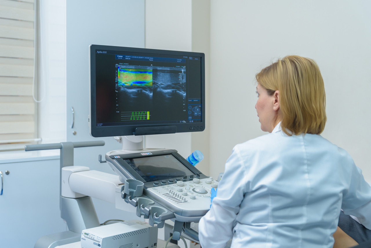
Ultrasound examination of the pelvic organs holds a paramount position among all diagnostic methods due to its high informativeness, non-invasiveness, and the ability to repeatedly conduct examinations dynamically without harming the patient's health. The high visualization capability of the equipment expands the possibilities for detecting pathologies and provides a more accurate picture of tissue microstructures, thereby enhancing the effectiveness of early-stage diagnosis and treatment.
Pelvic ultrasound is a non-invasive and safe diagnostic method that allows obtaining information about the condition of internal organs. Pelvic ultrasound helps detect many diseases at an early stage.
Pelvic ultrasound is recommended for various medical conditions. The main indications include:
- Lower abdominal pain
- Menstrual cycle disorders
- Heavy or painful periods
- Suspected tumors, cysts, or fibroids
- Infertility issues
- Inflammatory processes in the pelvic organs
- Post-surgical condition monitoring
Regular pelvic ultrasound is also recommended as a preventive examination for women of all ages, especially during menopause.
Pelvic ultrasound allows for a detailed examination of the following organs:
- Uterus
- Ovaries
- Fallopian tubes
- Bladder
Preparation for pelvic ultrasound depends on the method of examination. If the ultrasound is performed through the abdominal wall (transabdominal ultrasound), it is necessary to drink 1-1.5 liters of water before the examination to fill the bladder. This will improve the visualization of the organs. If the ultrasound is performed using a vaginal probe (transvaginal ultrasound), preparation includes standard hygiene procedures. It is recommended to avoid foods that cause gas before the examination, as this may reduce the image quality.
The most optimal time for the examination is the 5th to 7th day of the menstrual cycle, immediately after its completion. During this period, the uterine lining (endometrium) is thin, which allows for better visualization of the organs' structure and the detection of potential pathologies. However, depending on the purpose of the examination, the doctor may recommend performing the ultrasound on other days.
Nairi Medical Center offers high-quality diagnostic services. Our center employs experienced specialists, ensuring diagnostic accuracy and patient comfort. Schedule a pelvic ultrasound in Yerevan at Nairi Medical Center by calling 8900 or visiting us at Paronyan 21.
Author:

Updated:
31 July 2025, 09:47Our certificates


List of services
- ▸ Plasmapheresis
- ▸ “Longevity Nairi”
- ▸ Laboratory Diagnosis
- ▸ Computed Tomography
- ▸ Magnetic Resonance Imaging
- ▸ Mammography
- ▸ X-Ray Diagnostics
- ▸ Ultrasound Diagnostics
- ▸ Endoscopic Diagnosis
- ▸ Functional diagnostics
- ▸ Electroneuromyography (ENMG)
- ▸ Electroencephalography (EEG)
- ▸ Vascular Ultrasound or Duplex Scan
- ▸ Kidney CT Scan
- ▸ Ultrasound (US) of the Lower Limbs
- ▸ Lymph Node Ultrasound (USG)
- ▸ CT Scan of the Heart
- ▸ CT Scan of the Liver
- ▸ Breast Ultrasound
- ▸ Pelvic Ultrasound
- ▸ Thyroid ultrasound
- ▸ Kidney Ultrasound
- ▸ Liver Ultrasound
- ▸ Ultrasound of the ovaries
- ▸ Ultrasound of the heart
- ▸ Abdominal ultrasound
- ▸ Ultrasound during pregnancy
- ▸ CT of paranasal sinuses
- ▸ Computed tomography of the chest
- ▸ Head/brain computed tomography
- ▸ Abdominal computed tomography
- ▸ Spinal MRI
- ▸ Joint MRI
- ▸ Brain MRI


