

Magnetic Resonance Imaging
Magnetic resonance imaging is a diagnostic examination that does not use ionizing radiation and provides detailed images of internal organs, soft tissues, joints, the spine, brain, blood vessels, and other anatomical structures. MRI is used to assess a wide range of diseases and structural abnormalities.
MRI may be indicated for neurological symptoms, headaches, back pain, joint problems, injuries, as well as suspected tumours or vascular disorders.
At Nairi Medical Center, MRI examinations of various organs and systems are performed using modern Siemens MAGNETOM Sola and MAGNETOM Vida 3T scanners.
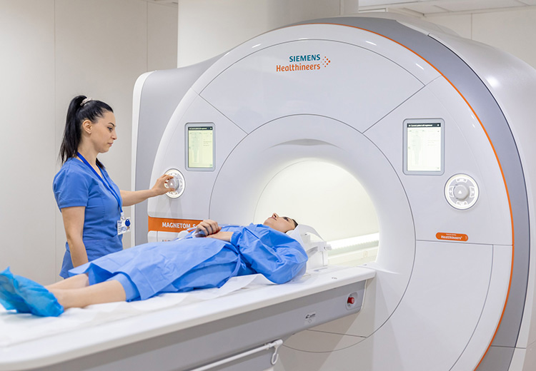
Magnetic Resonance Imaging (MRI) is an advanced diagnostic technique that uses magnetic fields and radio waves to obtain detailed images of internal organs without ionizing radiation.
MRI is widely used to diagnose and assess conditions affecting the brain, spine, joints, abdominal and pelvic organs, blood vessels, and soft tissues.
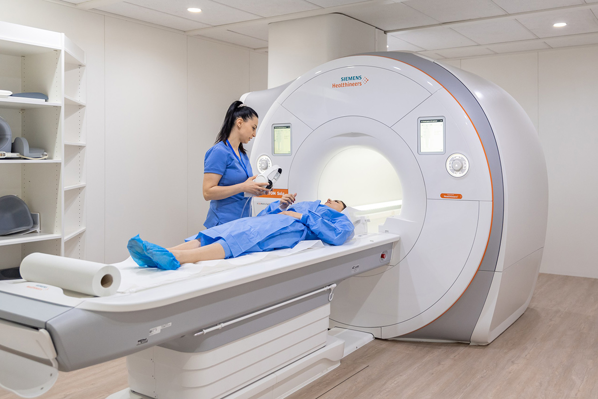
When MRI Is Recommended
Your doctor may refer you for an MRI scan to clarify or confirm a diagnosis.
MRI may be indicated in cases such as:
- Persistent or unexplained headaches unresponsive to standard treatment
- Neurological symptoms — seizures, numbness, weakness, dizziness, vision changes, or insomnia
- Vascular pathologies
- Head or neck injuries
- Chronic or severe back pain
- Joint injuries and degenerative disorders
- Suspected tumors or neoplasms in any organ
- Diseases of the abdominal or pelvic organs
Main Areas of MRI Diagnostics
Head and Neck: brain, pituitary gland, cranial nerves, soft tissues, and vessels.
Spine: cervical, thoracic, and lumbar–sacral regions with detailed visualization of discs, spinal cord, and nerves.
Chest: heart, major vessels, and mammary glands.
Abdomen and Retroperitoneum: liver, gallbladder, spleen, pancreas, kidneys, adrenal glands, intestines, and aorta.
Pelvis: urinary and reproductive organs — bladder, prostate, uterus, ovaries, and rectum.
Joints and Bones: all major joints — hip, knee, shoulder, elbow, wrist, sacroiliac, and temporomandibular.
Soft Tissues: inflammatory, traumatic, and tumor pathologies in any body area.
Vessels (MR Angiography): arteries and veins of the head, neck, abdomen, and limbs.
Equipment
Our center is equipped with next-generation MRI systems that enable high-quality imaging in minimal time.

MAGNETOM Sola
- BioMatrix Technology — automatically adapts to each patient’s anatomy, reducing artifacts and improving diagnostic accuracy.
- Turbo Suite — accelerates scans by up to 50%.
- Wide bore (70 cm), short magnet (145 cm), built-in lighting and music for patient comfort, especially in claustrophobic cases.
- Free-breathing capability — ideal for children, elderly, and respiratory patients.
- Artificial intelligence for automatic positioning and enhanced image quality.
- Suitable for patients with implants or other MRI limitations.
MAGNETOM Vida 3T
- High-speed, high-resolution imaging with Turbo Suite technology.
- GO Technologies streamline every step from positioning to analysis.
- Exceptional 3T image uniformity and reliable fat suppression — optimal for abdominal and peripheral scans.
- Advanced clinical applications — whole-body MRI, cardiac imaging with free breathing (GRASP-VIBE), quantitative diagnostics, and radiotherapy planning.
Our MRI department operates 24/7 for your convenience.
+374 (10) 53 69 33 | +374 (99) 99 16 16
21 Paronyan Street, Yerevan
Conditions Diagnosed by MRI
- Congenital anomalies and brain injuries
- Inflammatory and vascular brain diseases
- Multiple sclerosis and other demyelinating disorders
- Paranasal sinus pathologies
- Tumors of any localization
- Spinal and spinal cord disorders
- Bone, joint, and soft-tissue pathologies
- Abdominal and pelvic organ diseases
List of diseases
Congenital anomalies and malformations of the brain
Post-traumatic brain injuries
Multiple sclerosis
Inflammatory and infectious diseases of the brain
Cerebrovascular diseases
Migraine
Oncological diseases of any localizatio
Inflammations of the paranasal sinuses
Pathology of the spine and spinal cord
Pathology of the bones and joints
Injuries of bones, soft tissues, and internal organs
Pathology of the abdominal cavity and pelvis


News
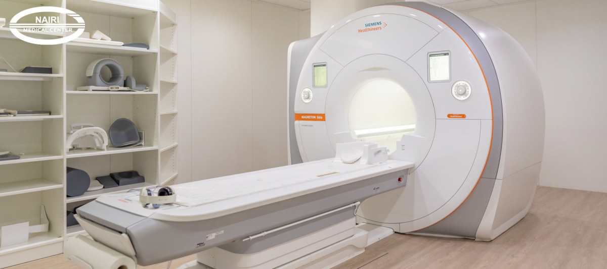
Nairi MC purchased an MRI MAGNETOM Sola with a power of 1.5 Tesla
Nairi Medical Center is equipped with heavy-duty equipment of the latest generation, which is the only one in Armenia
MAGNETOM Sola 1.5 Tesla MRI from Siemens Healthineers, equipped with artificial intelligence, helps carry out large-scale studies in the shortest possible time.

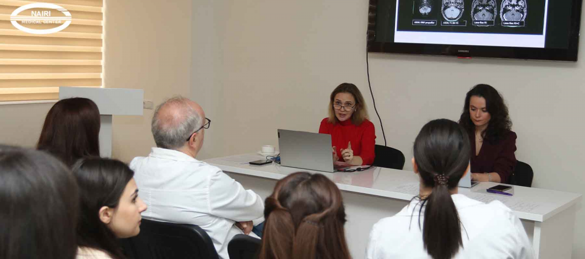

Representatives of Nairi and Erebuni Medical Centers participated in the Annual Congress of the Radiological Society of North America
The 109th Annual Scientific Congress of the Radiological Society of North America RSNA was held in Chicago.
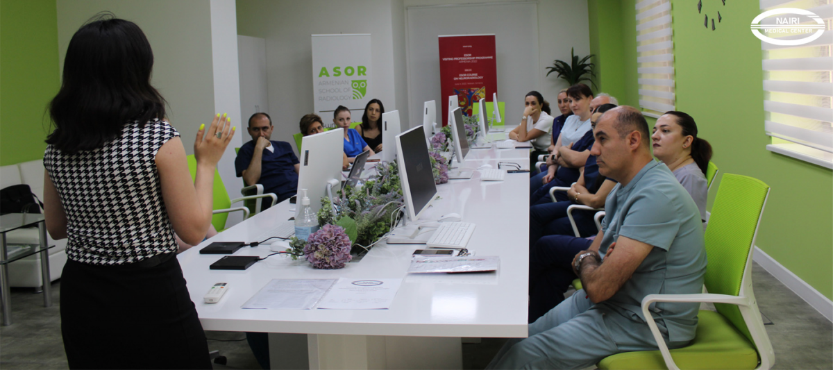
Within the introduction of the ISO 9001:2015 international quality management standard and the authoritative Joint Commission International (JCI) accreditation process
Within the introduction of the ISO 9001:2015 international quality management standard and the authoritative Joint Commission International (JCI) accreditation process, the awareness courses continue in the "Nairi" Medical Center



Frequently Asked Questions
An MRI scan can be booked in advance through the medical center’s registration department. During booking, the type of examination, preparation requirements, previous examination results, and available appointment times are usually clarified. If contrast-enhanced MRI is planned, additional questions may be asked about the patient’s health and coexisting conditions.
In many cases, a referral for MRI is advisable because it helps clarify the purpose of the examination and select the correct area to be scanned. Administrative requirements should preferably be clarified when booking the appointment.
The duration of an MRI scan depends on the area being examined and the imaging protocol. It usually takes about 15–40 minutes, but in some cases it may take longer if several areas are examined or contrast material is used. It is important to remain still during the scan, as movement can affect image quality.
MRI can also be performed in children when medically indicated. The possibility of the examination depends on the child’s age, overall condition, ability to remain still, and the type of scan. Since MRI requires remaining motionless for a certain period, the approach for younger children is determined individually by the physician.
In many cases, no special dietary restrictions are required before MRI, although this depends on the type of examination. Some scans, particularly MRI of the abdomen or pelvic organs, may require preparation in advance. If contrast material is planned, preparation instructions may also differ.
How often can MRI be performed?
MRI images and the medical report are usually provided to the patient the day after the examination, while in urgent situations they may be available on the same day.
MRI results are interpreted and reported by a radiologist. The radiologist evaluates the images, describes any detected abnormalities, and prepares a report. The final clinical interpretation is usually made by the treating physician, who considers the MRI findings together with the patient’s symptoms, physical examination, and other test results.
The cost of MRI usually includes the scan itself and the radiology report. However, the final scope of the service may vary depending on the examination format. If contrast material is used, additional imaging sequences are performed, or special protocols are required, these may be charged separately. It is advisable to clarify the cost structure when booking.
The possibility of having an MRI without a prior appointment depends on appointment availability and the medical center’s schedule. Because some examinations require special preparation or advance planning for contrast administration, booking in advance is generally recommended. This makes it possible to choose a suitable time and receive the necessary preparation instructions beforehand.
Author:

Updated:
10 August 2025, 18:43Our certificates



List of services
- ▸ Plasmapheresis
- ▸ “Longevity Nairi”
- ▸ Laboratory Diagnosis
- ▸ Computed Tomography
- ▸ Magnetic Resonance Imaging
- ▸ Mammography
- ▸ X-Ray Diagnostics
- ▸ Ultrasound Diagnostics
- ▸ Endoscopic Diagnosis
- ▸ Functional diagnostics
- ▸ Electroneuromyography (ENMG)
- ▸ Electroencephalography (EEG)
- ▸ Vascular Ultrasound or Duplex Scan
- ▸ Kidney CT Scan
- ▸ Ultrasound (US) of the Lower Limbs
- ▸ Lymph Node Ultrasound (USG)
- ▸ CT Scan of the Heart
- ▸ CT Scan of the Liver
- ▸ Breast Ultrasound
- ▸ Pelvic Ultrasound
- ▸ Thyroid ultrasound
- ▸ Kidney Ultrasound
- ▸ Liver Ultrasound
- ▸ Ultrasound of the ovaries
- ▸ Ultrasound of the heart
- ▸ Abdominal ultrasound
- ▸ Ultrasound during pregnancy
- ▸ CT of paranasal sinuses
- ▸ Computed tomography of the chest
- ▸ Head/brain computed tomography
- ▸ Abdominal computed tomography
- ▸ Spinal MRI
- ▸ Joint MRI
- ▸ Brain MRI


