Joint MRI
Magnetic resonance imaging (MRI) is used to perform diagnostic studies of various organ systems of the body without X-ray radiation, including magnetic resonance angiography (MRA) and whole-body MRI with three-dimensional imaging.
Magnetic resonance imaging (MRI) of the joints is a highly precise examination method that allows getting a detailed picture of the joint structure, without the use of X-ray radiation. This method is one of the most important components in diagnosing various diseases and injuries of the musculoskeletal system.
In what cases is it performed?
Joint MRI is prescribed when suspecting a number of pathologies, such as injuries, inflammations, deformations, or tumors in the joint area. An MRI may be ordered by an orthopedic surgeon, rheumatologist, traumatologist, or general surgeon.
What examinations do we perform?
MRI of the knee
Allows a detailed assessment of the structure of the knee joint, detecting injuries to the meniscus, cartilage, ligaments, and tumor formations.
MRI of the shoulder
Examines the structures of the shoulder joint, including the joint capsule, soft tissue, and tendons. Helps to identify arthritis, injuries, and other disorders.
MRI of the hip joint
Provides an image of the hip joint, while detecting any degenerative changes, arthritis, and even oncological problems at early stages.
MRI of other joints
In addition to the above-mentioned procedures, other joints (elbow joint, wrist joint, etc.) can also be examined, depending on the medical requirement.
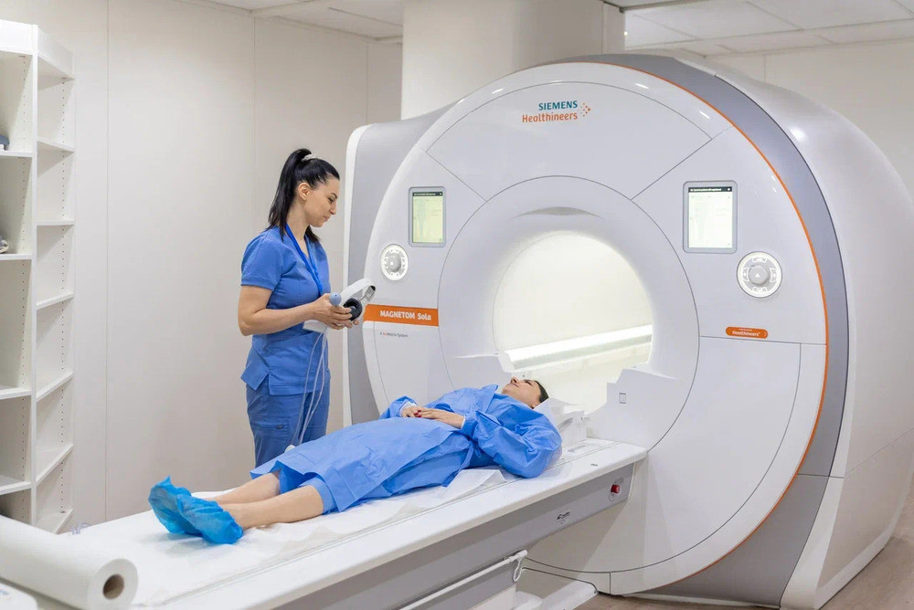
Our equipment; Magnetom Vida 3T & Magnetom Sola 1.5 T
We use advanced equipment to perform MRI examinations. Magnetom Vida 3T provides high image definition thanks to its 3 Tesla magnetic field. Magnetom Sola 1.5 T, with a magnetic field of 1.5 Tesla, provides high-quality results and a comfortable procedure.
Characteristics of the equipment
Magnetom Vida 3T provides fast scanning, and the ability to use innovative technologies, such as functional MRI.
Magnetom Sola 1.5 T provides a wide range of convenient applications.
Where to do joint MRI in Yerevan - Nairi Medical Center
You can undergo a high-quality MRI examination of the joints at Nairi Medical Center in Yerevan. Our highly qualified specialists work with modern equipment, guaranteeing accurate results and a caring approach to each patient.
Author:

Updated:
31 July 2025, 11:20Our certificates


List of services
- ▸ Plasmapheresis
- ▸ “Longevity Nairi”
- ▸ Laboratory Diagnosis
- ▸ Computed Tomography
- ▸ Magnetic Resonance Imaging
- ▸ Mammography
- ▸ X-Ray Diagnostics
- ▸ Ultrasound Diagnostics
- ▸ Endoscopic Diagnosis
- ▸ Functional diagnostics
- ▸ Electroneuromyography (ENMG)
- ▸ Electroencephalography (EEG)
- ▸ Vascular Ultrasound or Duplex Scan
- ▸ Kidney CT Scan
- ▸ Ultrasound (US) of the Lower Limbs
- ▸ Lymph Node Ultrasound (USG)
- ▸ CT Scan of the Heart
- ▸ CT Scan of the Liver
- ▸ Breast Ultrasound
- ▸ Pelvic Ultrasound
- ▸ Thyroid ultrasound
- ▸ Kidney Ultrasound
- ▸ Liver Ultrasound
- ▸ Ultrasound of the ovaries
- ▸ Ultrasound of the heart
- ▸ Abdominal ultrasound
- ▸ Ultrasound during pregnancy
- ▸ CT of paranasal sinuses
- ▸ Computed tomography of the chest
- ▸ Head/brain computed tomography
- ▸ Abdominal computed tomography
- ▸ Spinal MRI
- ▸ Joint MRI
- ▸ Brain MRI


