

Ultrasound Diagnostics
Ultrasound examination is a safe diagnostic method that does not involve radiation exposure. It allows physicians to assess the condition of internal organs, soft tissues, blood vessels, as well as fetal development during pregnancy. The method is used to examine the abdominal organs, pelvic organs, thyroid gland, breasts, heart, joints, and other anatomical structures.
Ultrasound is an effective tool for evaluating various organs and body systems. It may be recommended in cases of pain, swelling, masses, urinary disorders, menstrual irregularities, and for pregnancy monitoring.
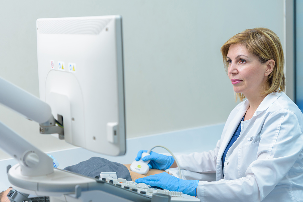
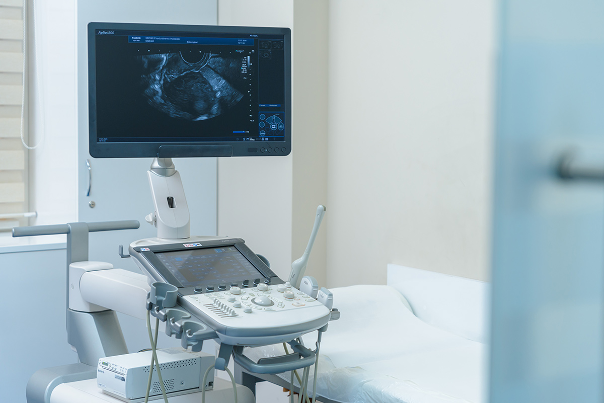
At Nairi Medical Center, ultrasound examinations of various organs and systems are performed using modern Canon Aplio I-Series equipment.
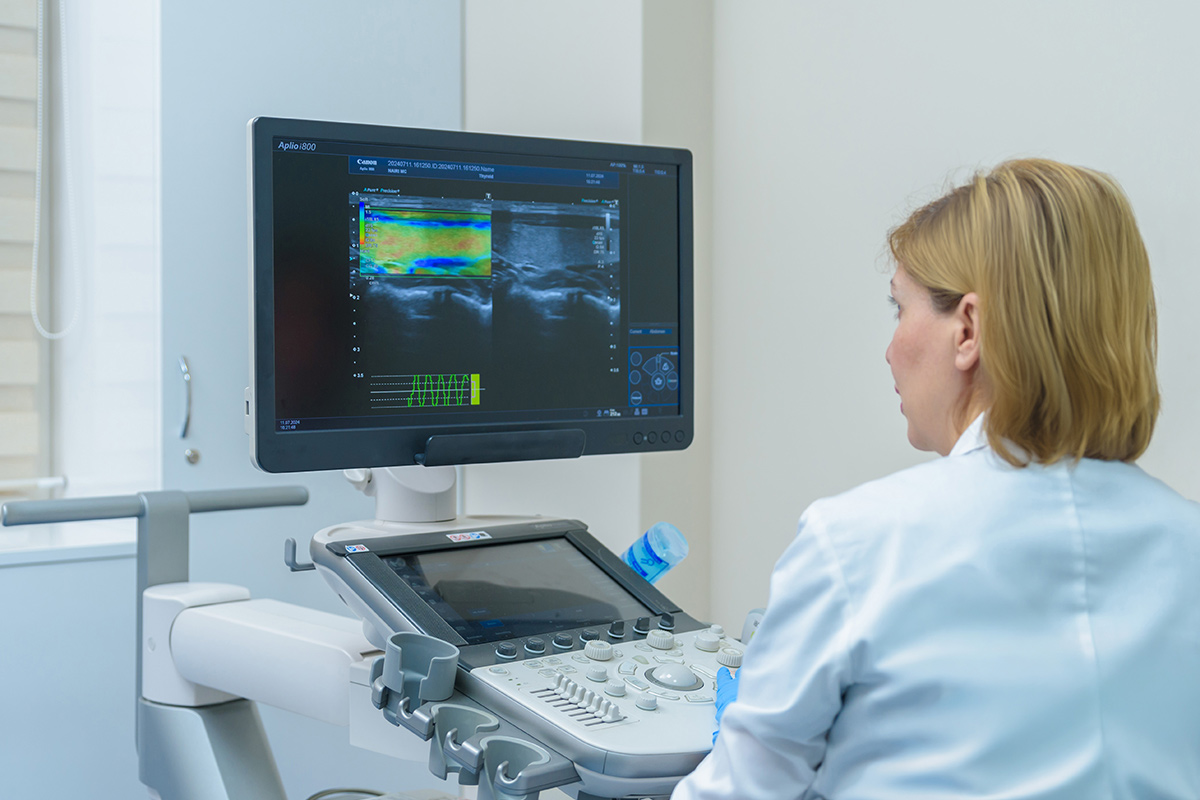
Ultrasound (US) is an important diagnostic method that allows doctors to obtain detailed images of a patient's internal organs and tissues without radiation exposure.
At the "Nairi" Medical Center, all medical examinations of organ systems are conducted:
- Abdominal organs
- Kidneys, adrenal glands
- Liver
- Ovaries
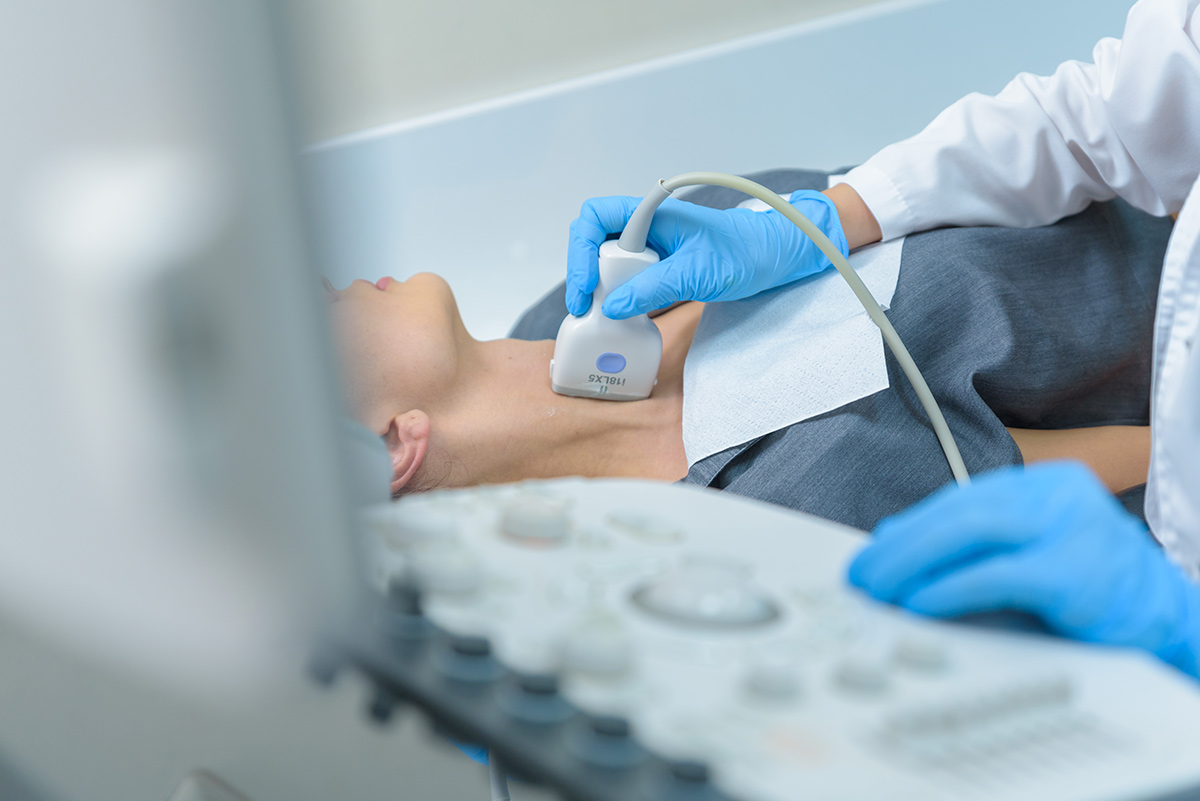
- Thyroid gland
- Lymph nodes and salivary glands
- Breast
- Pelvic organs
- Pelvic organs
- Soft tissues and joints
- Triplex scanning of major vessels and internal organ vessels
- Pregnancy
- Dopplerography of uterine and fetal blood circulation
- Echocardiography and stress echocardiography
- Obtaining comprehensive images of the fetus and any organ
- Lower Limbs
- Vascular (duplex scan)
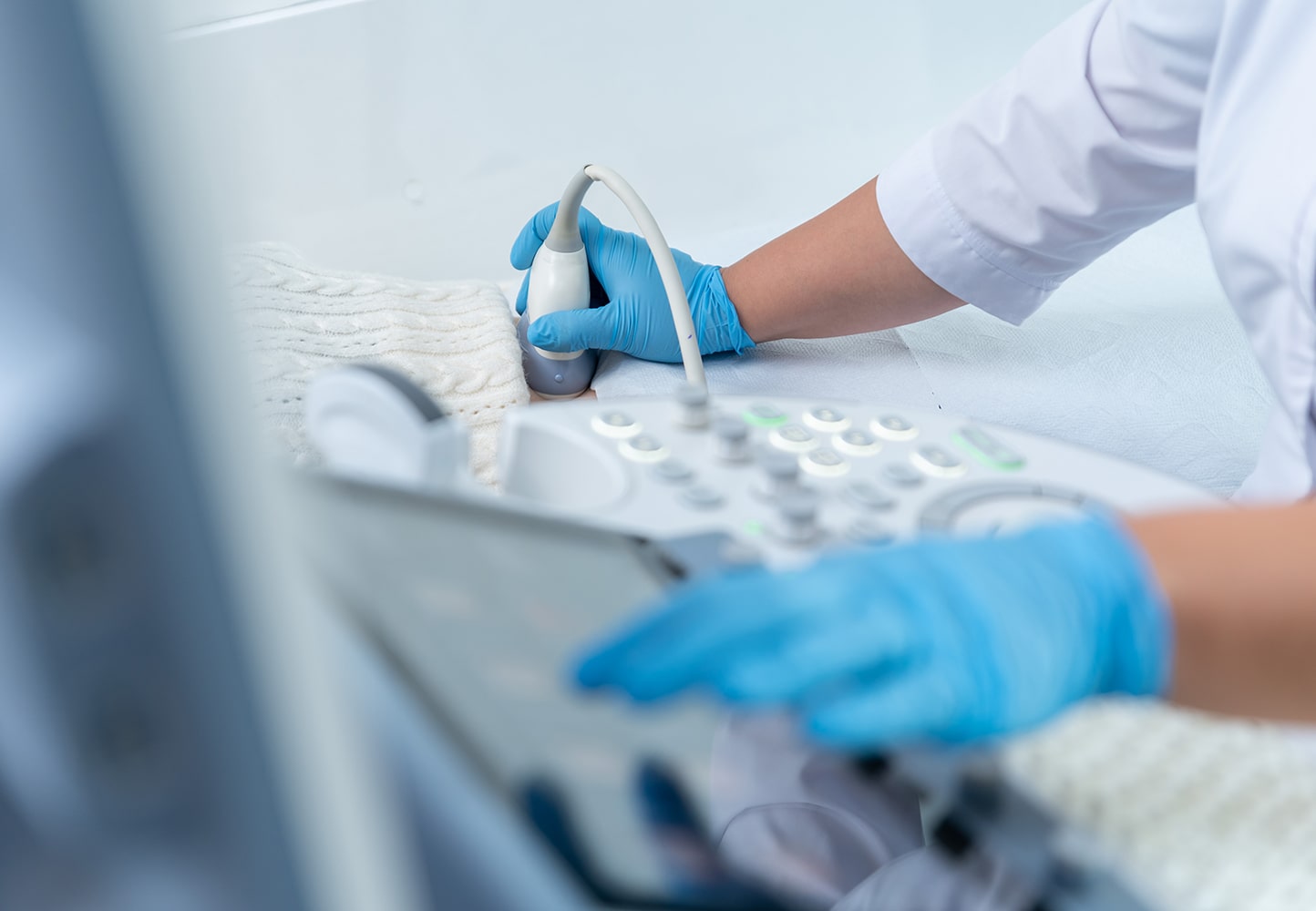
Indications for a Visit
A medical examination is necessary if the following symptoms are present:
- Abdominal pain: If you have frequent abdominal pain, an ultrasound can help identify issues such as problems with the liver, gallbladder, kidneys, or female reproductive organs.
- Swelling: This may be a result of issues with the heart, kidneys, or blood vessels.
- Urination problems
- Formation of new lumps: Any new lumps, thickenings, or cysts under the skin can be examined with ultrasound to determine their nature.
Our Equipment
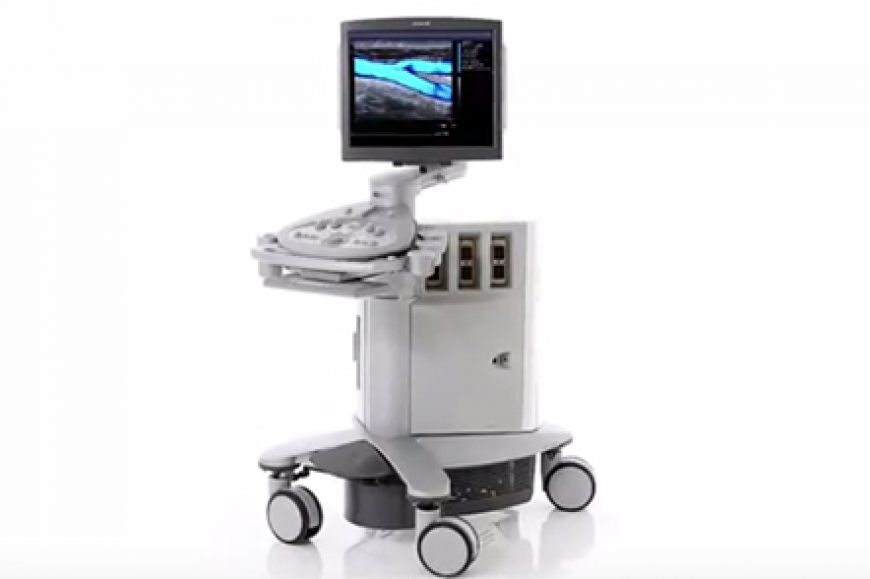
The "Nairi" Medical Center uses the best equipment, Canon Aplio I-Series, for medical examinations. This ultrasound imaging system is known for its advanced features and high-quality imaging capabilities.
The "Nairi" Medical Center is equipped with the latest medical equipment and provides the best medical services in Yerevan. To schedule an ultrasound examination, you can call +37410536933 and +37499991616 or visit 21 Paronyan Street.
List of diseases
Gynecological pathology
Pathologies of the head and neck region
Endocrine diseases
Pathologies of the digestive system and abdominal cavity
Pathologies of the respiratory system and thoracic cavity
Tumors
Pathologies of the genitourinary system
Heart and blood vessels
Ear, throat, nose
Pathologies of the bones and joints
Doctors







News


Frequently Asked Questions
The duration depends on the area being examined and the purpose of the study. In most cases, it takes approximately 10 to 30 minutes. If several organs are being assessed or Doppler ultrasound is required, the total time may be longer.
Unlike X-ray imaging, ultrasound uses high-frequency sound waves. It is a non-invasive and safe method. It is widely used for patients of all age groups, including children and pregnant women, when medically indicated.
Ultrasound can be performed as often as required by the clinical situation and the physician’s assessment. Since the method does not involve radiation exposure, it is commonly used for follow-up and dynamic monitoring. The frequency depends on symptoms, diagnosis, treatment progress, and the need to monitor a specific organ or system.
Yes, ultrasound is widely used in pediatric practice. It is performed to assess the abdominal organs, kidneys, thyroid gland, joints, soft tissues, and, in newborns, the brain as well. Because it is safe and non-invasive, it is often chosen as a primary or follow-up diagnostic method for children.
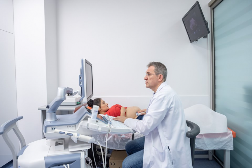
Yes, ultrasound is routinely used during pregnancy to evaluate fetal growth, the placenta, amniotic fluid volume, and blood circulation. During pregnancy, scheduled and additional ultrasound examinations may be performed according to the obstetrician-gynecologist’s recommendation. The type and frequency of examinations depend on the gestational age and clinical situation.
This depends on the type of examination. Some ultrasound studies do not require dietary restrictions, while for abdominal ultrasound patients are often advised not to eat for several hours beforehand. This helps provide clearer visualization of the liver, gallbladder, pancreas, and other organs. Preparation instructions should be confirmed at the time of booking.
This also depends on the area being examined. In some cases, there are no restrictions, while for pelvic organ or bladder ultrasound, a full bladder may be required. Therefore, it is important to know in advance which type of ultrasound is planned and what preparation is needed.
In many cases, results are available immediately after the examination or shortly afterwards. Since the physician evaluates the images during the procedure, the report is often prepared during the same visit. If the examination is extensive or requires a more detailed description, additional time may be needed.
The results are evaluated and described by a qualified specialist. The report includes organ size, tissue structure, echogenicity, the presence of masses or other changes, and a medical conclusion. Final clinical interpretation is provided by the treating physician, who correlates the ultrasound findings with the patient’s symptoms, physical examination, and other test results.
Author:

Updated:
25 October 2025, 05:45Our certificates



List of services
- ▸ Plasmapheresis
- ▸ “Longevity Nairi”
- ▸ Laboratory Diagnosis
- ▸ Computed Tomography
- ▸ Magnetic Resonance Imaging
- ▸ Mammography
- ▸ X-Ray Diagnostics
- ▸ Ultrasound Diagnostics
- ▸ Endoscopic Diagnosis
- ▸ Functional diagnostics
- ▸ Electroneuromyography (ENMG)
- ▸ Electroencephalography (EEG)
- ▸ Vascular Ultrasound or Duplex Scan
- ▸ Kidney CT Scan
- ▸ Ultrasound (US) of the Lower Limbs
- ▸ Lymph Node Ultrasound (USG)
- ▸ CT Scan of the Heart
- ▸ CT Scan of the Liver
- ▸ Breast Ultrasound
- ▸ Pelvic Ultrasound
- ▸ Thyroid ultrasound
- ▸ Kidney Ultrasound
- ▸ Liver Ultrasound
- ▸ Ultrasound of the ovaries
- ▸ Ultrasound of the heart
- ▸ Abdominal ultrasound
- ▸ Ultrasound during pregnancy
- ▸ CT of paranasal sinuses
- ▸ Computed tomography of the chest
- ▸ Head/brain computed tomography
- ▸ Abdominal computed tomography
- ▸ Spinal MRI
- ▸ Joint MRI
- ▸ Brain MRI


