

Electroencephalography (EEG)
Electroencephalography is an instrumental examination used to record the electrical activity of the brain. It is used to assess various neurological conditions, including disorders of consciousness, seizure-related conditions, functional changes in the brain, and a range of other problems.
EEG may be indicated in cases of seizures, fainting, episodes of loss of consciousness, certain sleep disorders, for assessment of brain function, as well as for other neurological complaints. At Nairi Medical Center, electroencephalography is performed according to clinical indications.
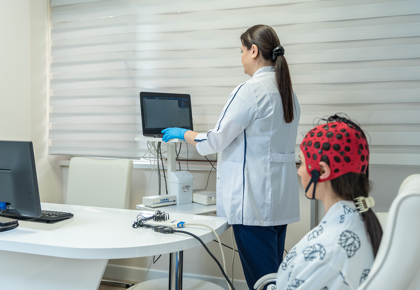
What is EEG
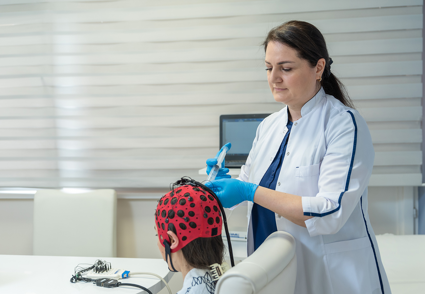
Electroencephalography (EEG) is a non-invasive method for studying the brain’s bioelectrical activity. It is based on recording electrical signals generated by neurons in the brain. During the procedure, special electrodes are placed on the scalp and connected to a device that records brain activity at different time intervals.
This method enables physicians to evaluate the functional state of the brain and detect various neurological disorders. EEG is safe and painless.
When EEG is Indicated
EEG is widely used in neurology and neurosurgery. It is indicated in cases of:
- diagnosis of epilepsy and clarification of seizure characteristics
- investigation of the causes of altered consciousness (dizziness, fainting, loss of consciousness)
- diagnosis of inflammatory brain diseases (encephalitis, meningoencephalitis)
- detection of cerebrovascular pathologies and monitoring recovery after stroke
- assessment of brain tumors or injuries
- evaluation of sleep disorders (sleep apnea, insomnia, etc.)
- monitoring treatment effectiveness in patients with epilepsy or other neurological conditions
EEG is also frequently performed prior to surgery to assess the brain’s functional state.
How EEG is Performed
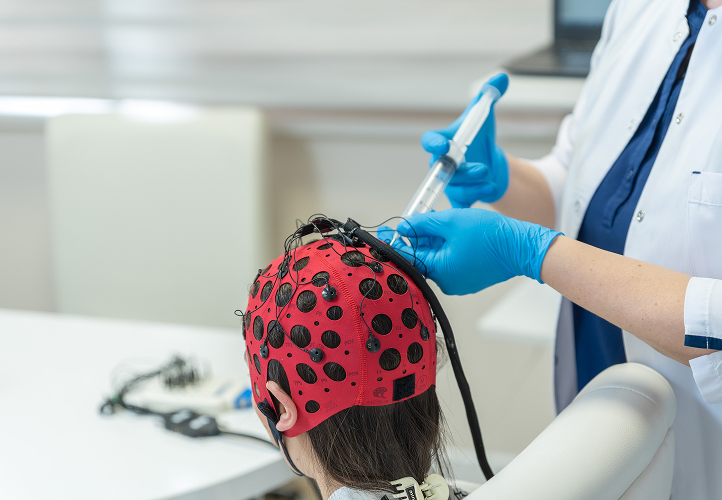
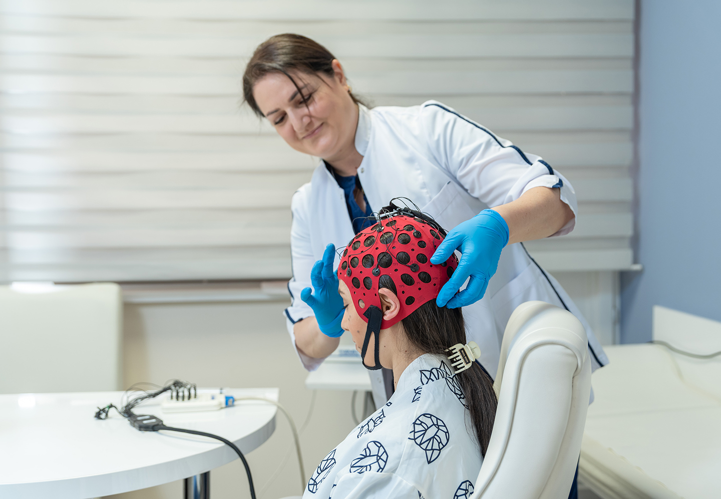
The procedure is carried out using specialized equipment — an electroencephalograph. The patient sits or lies comfortably, while small electrodes are placed on the scalp and fixed with conductive gel. These electrodes record the brain’s electrical activity and transmit it to a computer.
EEG can be performed in different durations: approximately 30 minutes, 1 hour, 2 hours, 24 hours, as well as EEG with sleep deprivation (1–2 hours). In some cases, additional functional tests — such as photic stimulation or hyperventilation — are applied to help reveal hidden abnormalities.
EEG is completely painless and has no side effects. After the procedure, the patient can immediately return to normal daily activities.
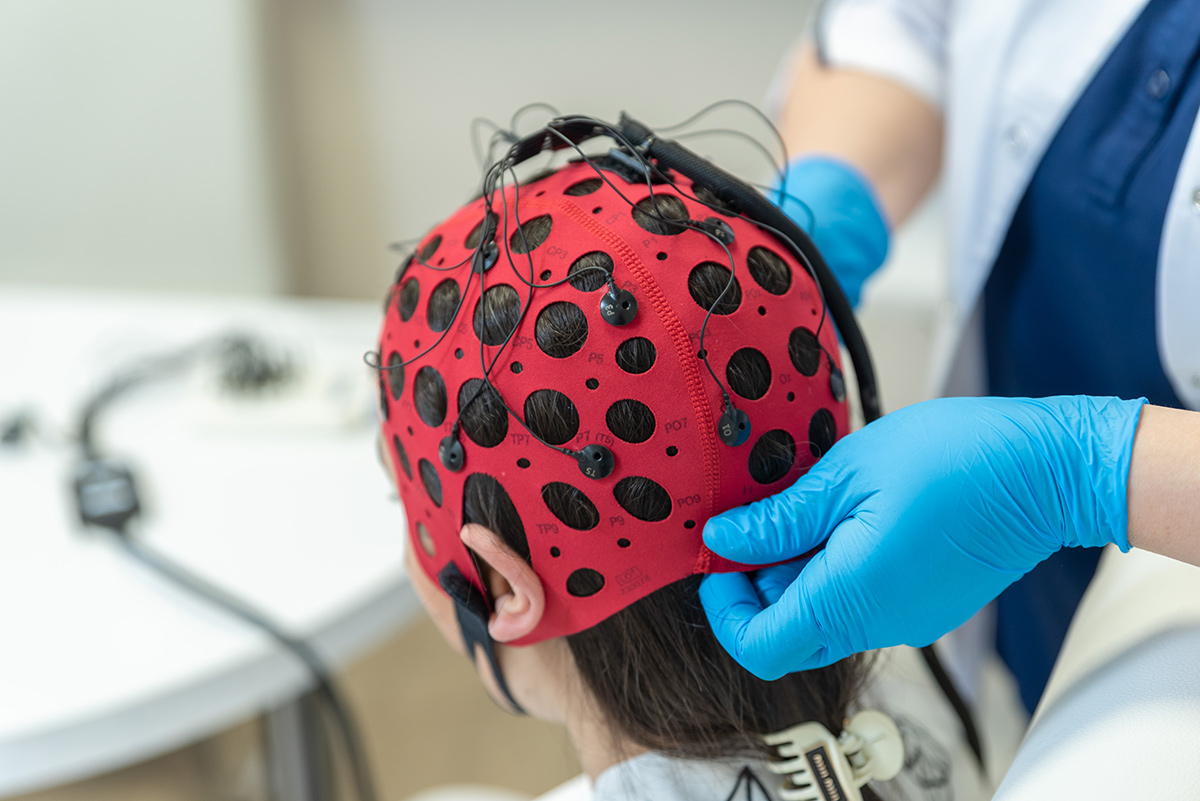
What EEG Shows
EEG reflects the electrical activity of different brain regions in the form of waves. By analyzing them, the neurologist can:
- detect foci of epileptic activity
- assess the overall level of bioelectrical activity
- monitor the effectiveness of drug therapy
- identify various organic or functional disorders
- evaluate the brain’s response to stimuli (light, sound, hyperventilation)
The results are interpreted by the physician and play an important role in diagnosis and treatment planning.
Where to Perform EEG in Yerevan
At Nairi Medical Center, EEG is performed with modern equipment by experienced neurologists. The center provides:
- high-precision devices for detailed brain activity recording
- comfortable conditions and individualized care
- possibility of conducting EEG in both adults and children
- professional result interpretation and consultation
EEG at Nairi Medical Center is completely safe, complies with international standards, and offers high diagnostic value.
To make an appointment, please call 89 00 or visit us at 21 Paronyan Street.



Frequently Asked Questions
The duration of electroencephalography depends on the type and purpose of the examination. An EEG may last 20 minutes, 1 hour, 2 hours, or 24 hours, depending on the type of recording prescribed.
Yes, electroencephalography can be performed in children; however, pediatric EEG examinations are not provided at Nairi Medical Center.
Electroencephalography results are often available on the same day or, depending on the duration and volume of the recording, within 1–2 days.
During a standard EEG, the brain’s electrical activity is recorded at rest. An EEG with activation procedures includes additional tests such as photic stimulation or deep breathing. These procedures may help assess certain functional changes more clearly and reveal abnormalities that may not be evident during a standard recording.
Electroencephalography can be performed during pregnancy when medically indicated. The appropriateness of the examination is determined by the physician, taking into account the patient’s symptoms, clinical condition, and other medical information.
MRI is an imaging examination that shows the structure of the brain, while electroencephalography assesses brain function by recording its electrical activity. These examinations do not replace one another and are used to evaluate different clinical problems.
Author:

Updated:
03 October 2025, 06:47Our certificates



List of services
- ▸ Thyroid Tests
- ▸ Cardiac Holter Monitoring
- ▸ Plasmapheresis
- ▸ “Longevity Nairi”
- ▸ Laboratory Diagnosis
- ▸ Computed Tomography
- ▸ Magnetic Resonance Imaging
- ▸ Mammography
- ▸ X-Ray Diagnostics
- ▸ Ultrasound Diagnostics
- ▸ Endoscopic Diagnosis
- ▸ Functional diagnostics
- ▸ Electroneuromyography (ENMG)
- ▸ Electroencephalography (EEG)
- ▸ Vascular Ultrasound or Duplex Scan
- ▸ Kidney CT Scan
- ▸ Ultrasound (US) of the Lower Limbs
- ▸ Lymph Node Ultrasound (USG)
- ▸ CT Scan of the Heart
- ▸ CT Scan of the Liver
- ▸ Breast Ultrasound
- ▸ Pelvic Ultrasound
- ▸ Thyroid ultrasound
- ▸ Kidney Ultrasound
- ▸ Liver Ultrasound
- ▸ Ultrasound of the ovaries
- ▸ Ultrasound of the heart
- ▸ Abdominal ultrasound
- ▸ Ultrasound during pregnancy
- ▸ CT of paranasal sinuses
- ▸ Computed tomography of the chest
- ▸ Head/brain computed tomography
- ▸ Abdominal computed tomography
- ▸ Spinal MRI
- ▸ Joint MRI
- ▸ Brain MRI


