
Ultrasound of the ovaries
Ultrasound examination of the ovaries holds a primary place among all diagnostic methods due to its high informativeness, non-invasiveness, and the ability to repeatedly conduct examinations in dynamics without harming the patient's health. The high visualization capability of the equipment enhances the ability to detect pathologies and provides a more accurate picture of the tissue microstructure, which allows for increased efficiency in diagnosis and treatment at the early stages of disease development.
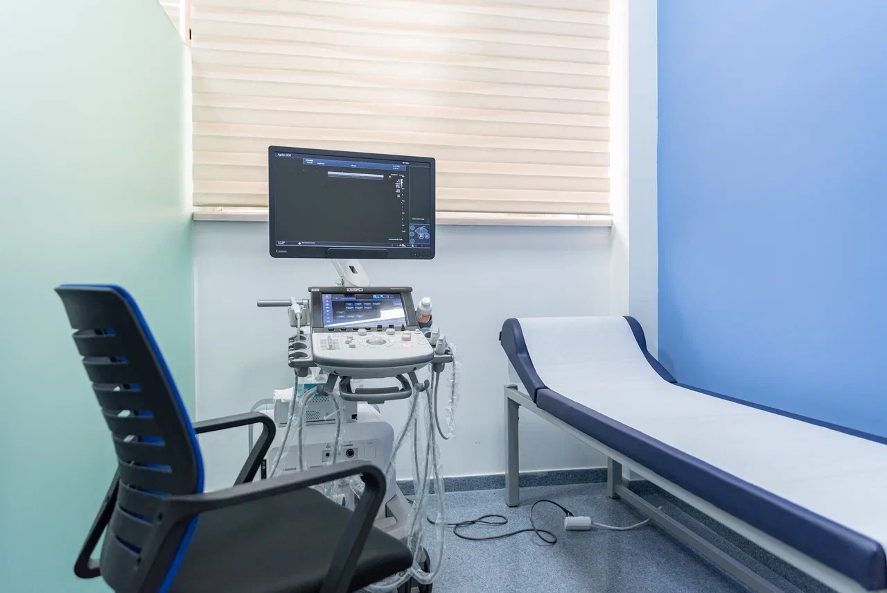
Ultrasound scan of the ovaries is an essential and frequently performed diagnostic examination. It allows one to visualize the ovaries, assess their size, structure, and detect any presence of cysts, tumors or other pathological changes. Ovarian ultrasound is a safe, non-invasive and informative diagnostic method. It doesn’t require the use of contrast has no harmful effects on the patient’s body.
There are two ways to perform ovarian ultrasound: transabdominal and transvaginal. Transabdominal ultrasound is performed through the abdominal wall and is suitable for preliminary examination as well as during pregnancy. Transvaginal ultrasound is performed using a special probe that is inserted into the vagina and allows a more detailed image of the ovaries and pelvic organs.
Indications for an examination
An ultrasound of the ovaries may be required in the following cases:
- Pain in the lower abdomen. Pain may indicate inflammation, cysts, or other ovarian abnormalities.
- Abnormalities of the menstruation: ultrasound helps to identify the causes of irregular or painful menstruation.
- Suspecting cysts or tumors: detection and monitoring of ovarian cysts or neoplasms.
- Infertility: assessment of the condition of the ovaries and follicles in case of problems with conception
- Monitoring of the treatment: monitoring the condition of the ovaries during hormone treatment or after surgery.
- Pregnancy planning: assessment of the reserve and condition of the ovaries before conception.
How to prepare for the examination?
The preparation for ovarian ultrasound depends on the method of examination:
Transabdominal ultrasound examination - the patient is recommended to drink 1-1.5 litres of water an hour before the examination without emptying the bladder.
Transvaginal ultrasound examination - no special preparation is required, but it is recommended to empty the bladder before the examination for patient’s improved comfort as well as image quality.
When to perform an examination?
The optimal time to perform an ovarian ultrasound is considered to be the first half of the menstrual cycle, usually from day 5 to day 10. During this period, the influence of hormones on the ovaries is minimal, which allows to obtain the most accurate data.
However, ultrasound can also be performed on other days of the cycle, which depends on the purpose of the examination.
To undergo an ovarian ultrasound examination in Yerevan, you can contact Nairi MC. In our medical center, the highly qualified specialists, with the use of modern ultrasound examination equipment, will ensure an accurate diagnosis as well as an individual approach to each patient.
Contact us by calling 8 900 or +37491555156 or visit us at Nairi MC, Paronyan Street 21.
Author:

Updated:
31 July 2025, 10:05Our certificates


List of services
- ▸ Plasmapheresis
- ▸ “Longevity Nairi”
- ▸ Laboratory Diagnosis
- ▸ Computed Tomography
- ▸ Magnetic Resonance Imaging
- ▸ Mammography
- ▸ X-Ray Diagnostics
- ▸ Ultrasound Diagnostics
- ▸ Endoscopic Diagnosis
- ▸ Functional diagnostics
- ▸ Electroneuromyography (ENMG)
- ▸ Electroencephalography (EEG)
- ▸ Vascular Ultrasound or Duplex Scan
- ▸ Kidney CT Scan
- ▸ Ultrasound (US) of the Lower Limbs
- ▸ Lymph Node Ultrasound (USG)
- ▸ CT Scan of the Heart
- ▸ CT Scan of the Liver
- ▸ Breast Ultrasound
- ▸ Pelvic Ultrasound
- ▸ Thyroid ultrasound
- ▸ Kidney Ultrasound
- ▸ Liver Ultrasound
- ▸ Ultrasound of the ovaries
- ▸ Ultrasound of the heart
- ▸ Abdominal ultrasound
- ▸ Ultrasound during pregnancy
- ▸ CT of paranasal sinuses
- ▸ Computed tomography of the chest
- ▸ Head/brain computed tomography
- ▸ Abdominal computed tomography
- ▸ Spinal MRI
- ▸ Joint MRI
- ▸ Brain MRI


