
Diagnosis and Treatment of Tear Duct Obstruction
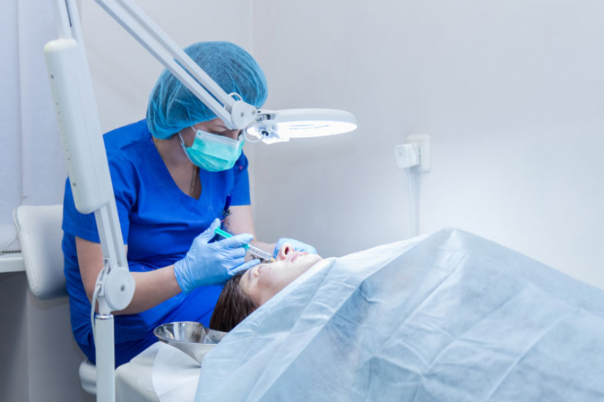
Normally, tears drain through small tear ducts that run from the eyes into the nasal cavity. When a tear duct becomes partially or completely blocked, normal tear drainage is disrupted. As a result, tears may accumulate in the lacrimal sac, leading to swelling, inflammation, and, in some cases, infection.
Tear duct obstruction occurs in approximately 6 out of every 100 newborns. In adults, it is less common and may develop as a result of age-related changes, trauma, or other underlying conditions.
One of the first signs of tear duct obstruction is excessive tearing, which may vary in severity. Symptoms can become more pronounced after upper respiratory infections. In adults, the obstruction is usually associated with an underlying condition or previous injury. It may develop due to thickening of the tissues lining the tear ducts, structural changes in the surrounding tissues or nasal bones, or as a complication following nasal surgery.
If an infection develops within the tear drainage system, redness or swelling may appear around the eyes or nose. Yellowish discharge may collect in the corners of the eyes, and the eyelids may stick together. In more severe cases, the infection may spread to the eyelids and surrounding tissues.
Symptoms may also become more noticeable in windy or cold weather, or when exposed to bright sunlight.
Diagnosis of tear duct obstruction is based on the patient’s medical history and a physical examination. Additional tests may be used to assess tear production and determine how effectively tears are draining from the eyes.
In Armenia, contrast dacryography, an advanced diagnostic method used to accurately determine the location and degree of tear duct narrowing, is available only at the Eye Clinic of Nairi Medical Center.
Treatment Options
If conservative treatment is not effective, interventional or surgical correction of the tear ducts may be recommended.
Probing involves passing a thin probe through the blocked tear duct to restore its patency. In children, probing successfully opens the duct in approximately 90 out of 100 cases. In adults, the procedure is generally less effective.
Intubation involves placing a silicone tube in the tear drainage system to create a pathway for tears to drain into the nasal cavity.
Creating an opening in the nasal bone may be performed to establish a wider drainage pathway into the nasal cavity.
Balloon dacryocystoplasty uses a small balloon attached to a probe to widen and open the blocked tear duct.
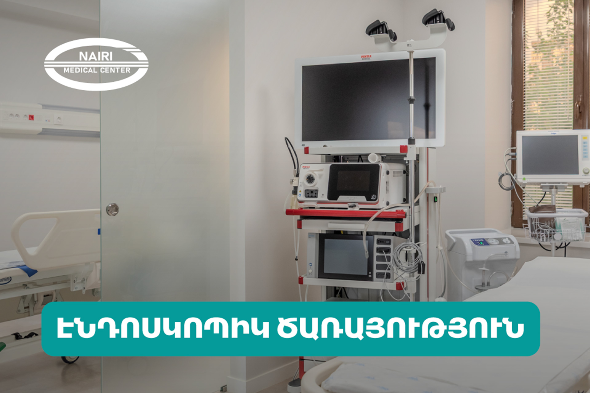
Endoscopic and laser dacryocystorhinostomy creates a new drainage pathway between the tear sac and the nasal cavity. The procedure is performed using endoscopic instruments and a flexible tube equipped with a small camera.
In adults, the type of surgical procedure selected for tear duct obstruction depends on the underlying cause and may include any of the methods described above.
For consultation and appointment scheduling, please contact: +374 10 502 522, +374 91 555 156, or 8900.
Watch more on our YouTube channel.